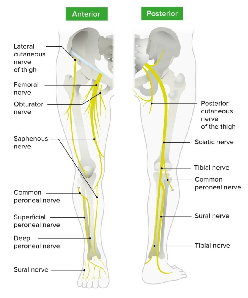
Understanding the nerves of the lower limb is crucial for healthcare professionals and students alike. The nerves of the lower limb are responsible for transmitting sensory and motor signals that control movement and sensation in the legs and feet. The nerves of the lower limb originate from the lumbar and sacral plexuses and branch out to innervate different muscles and skin regions.
One of the most effective ways to study and memorize the nerves of the lower limb is through a flow chart. A flow chart visually represents the pathway of each nerve, making it easier to understand their origins, branches, and functions.
Nerves Of Lower Limb Flow Chart
The Sciatic Nerve
The sciatic nerve is the largest nerve in the lower limb and is composed of two main branches: the tibial nerve and the common peroneal nerve. The sciatic nerve originates from the sacral plexus and runs down the back of the thigh, branching out to innervate the muscles of the thigh, leg, and foot. The tibial nerve supplies sensation to the sole of the foot, while the common peroneal nerve innervates the muscles that dorsiflex the foot.
Damage to the sciatic nerve can result in pain, numbness, and weakness in the lower limb. Understanding the pathway of the sciatic nerve through a flow chart can help healthcare professionals diagnose and treat nerve injuries more effectively.
The Femoral Nerve
The femoral nerve is another important nerve of the lower limb that originates from the lumbar plexus. It runs down the front of the thigh and innervates the quadriceps muscle, which is responsible for extending the knee. The femoral nerve also supplies sensation to the skin of the thigh and inner leg.
A flow chart detailing the pathway of the femoral nerve can help healthcare professionals pinpoint the location of nerve compression or injury, leading to more accurate treatment and rehabilitation strategies.
